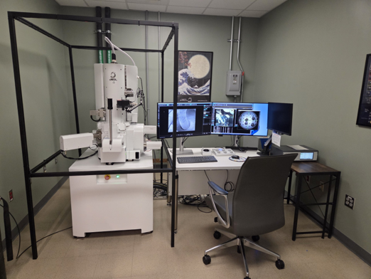
JEOL JSM-IT810(HL) FE-SEM at UAB HRIF.
The JEOL JSM-IT810(HL) is a high-resolution field emission scanning electron microscope (FE-SEM) located in the UAB High Resolution Imaging Facility (HRIF). The instrument combines a Schottky field emission electron source with a hybrid electromagnetic/electrostatic objective lens for detailed examination of specimen surface morphology and microstructure. The system supports imaging and elemental analysis for biological, materials science, and nanotechnology research.
The JSM-IT810(HL) is equipped with multiple secondary and backscattered electron detectors, a retractable low-vacuum system, and a JEOL energy-dispersive X-ray spectroscopy (EDS) detector for elemental characterization. Its five-axis motorized stage, integrated stage navigation system, and ZEROMAG functionality assist users in locating regions of interest and transitioning from optical survey images to electron microscopy.
Computer-controlled operation, automated alignment and focus functions, image acquisition, and montage tools support a range of analytical workflows. HRIF staff provide instrument access, training, and assistance with sample preparation and imaging.
Features:
- High-resolution FE-SEM imaging with manufacturer-specified resolution of 0.7 nm at 20 kV and 1.3 nm at 1 kV; analytical resolution of 3.0 nm at 15 kV (5 nA).
- Schottky in-lens thermal field emission electron gun with an accelerating voltage range of 0.01–30 kV.
- Hybrid electromagnetic/electrostatic objective lens for high-resolution imaging of a broad range of specimens, including magnetic materials.
- Gentle Beam specimen biasing and beam deceleration capability for low landing-energy imaging and reduced charging.
- Secondary and backscattered electron imaging using an Everhart–Thornley secondary electron detector, upper electron detector with energy filtering, and retractable scintillator backscattered electron detector.
- Retractable low-vacuum system with low-vacuum secondary electron detector to support imaging of specimens that are difficult to examine under high vacuum.
- JEOL EDS detector (60 mm² nominal active area) for elemental X-ray microanalysis.
- Five-axis motorized specimen stage with computer-eucentric tilt compensation and a 100 mm specimen exchange chamber.
- Stage Navigation System and ZEROMAG optical-to-SEM navigation for finding and revisiting regions of interest.
- NEOENGINE electron-optical control, Aperture Angle Control Lens, automated operating functions, and Smile View image management and analysis software.
- EMS combined sputtering and carbon coating system is available in HRIF to support sample preparation.